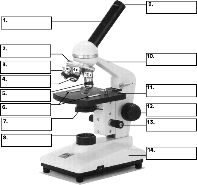
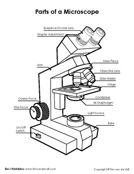
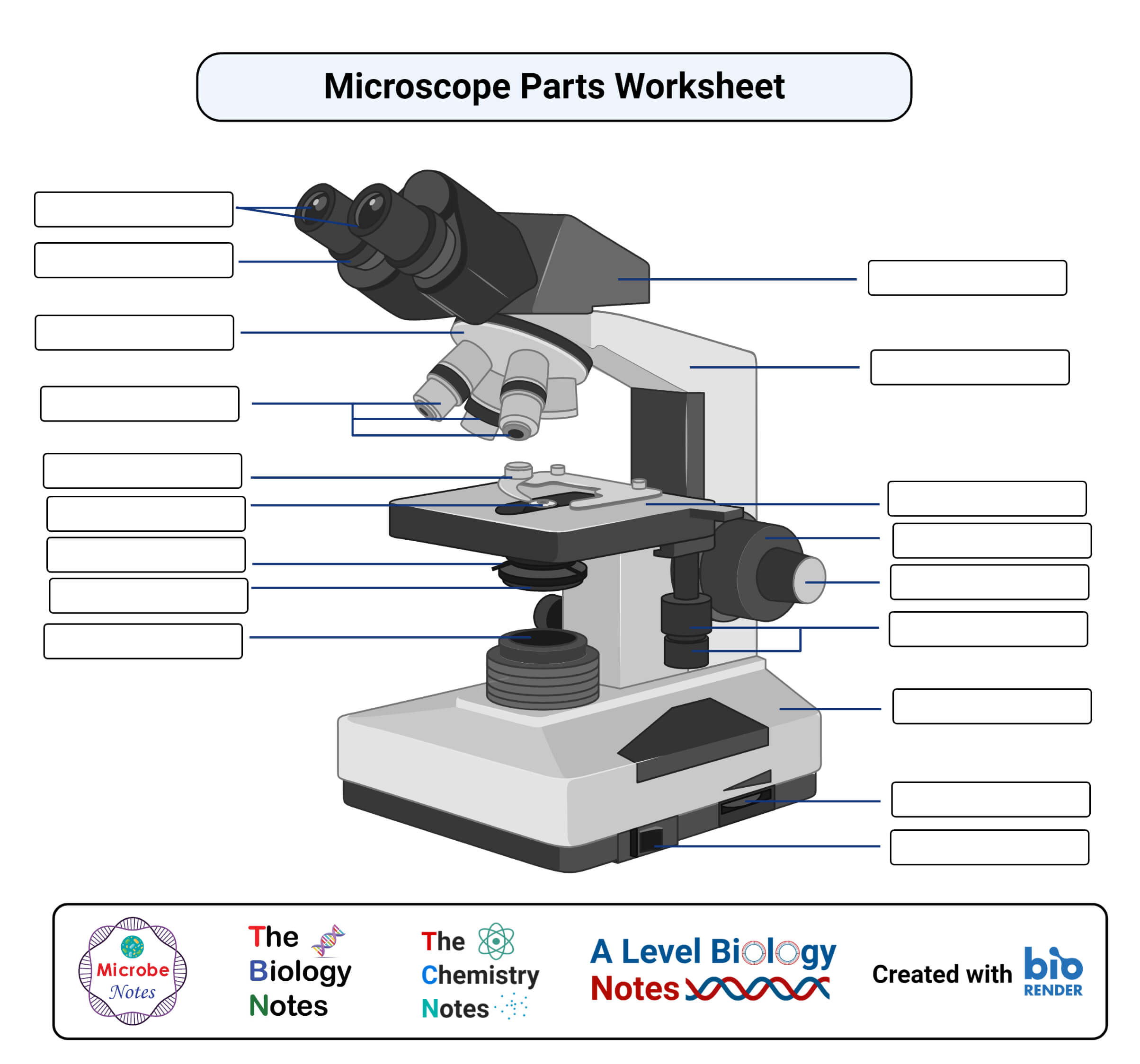
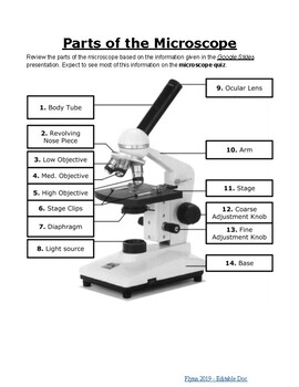
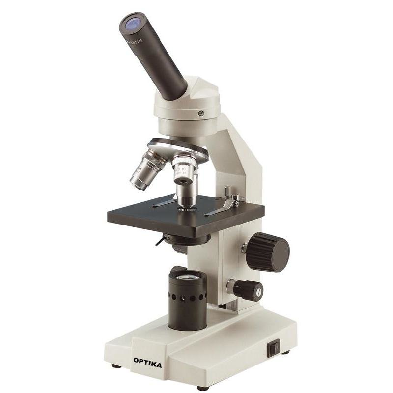
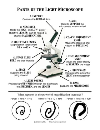
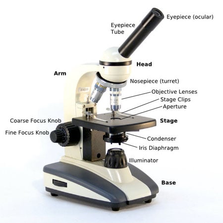
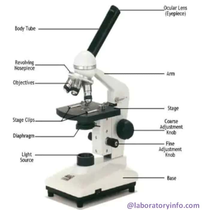
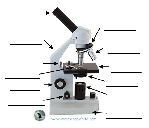
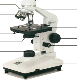
41 blank microscope diagram
Plant Cell: Definition, Types of Plant Cells and More - Embibe These differences can be clearly understood when the cells are examined under an electron microscope. Observe the labelled diagram of plant cell structure as given below: Are Plant Cells Prokaryotic or Eukaryotic? The cell is the basic structural and functional unit of life in all living organisms. The cells can be divided into two major groups ... Optical dispersion properties of topological photonic crystals using ... We developed a photonic band diagram microscope using hyperspectral Fourier image spectroscopy to investigate optical properties of topological photonic crystals. The band inversion near the Γ point was observed in the high-speed measurement.
Electron Microscope Principle, Uses, Types and Images (Labeled Diagram ... Ans: A light microscope has a low resolving power (0.25µm to 0.3µm) while the electron microscope has a resolution power about 250 times higher than the light microscope at about 0.001µm. Similarly, a light microscope has a magnification of 500X to 1500x while the electron microscope has a much higher magnification of 100,000X to 300,000X.
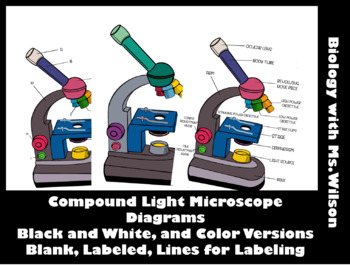
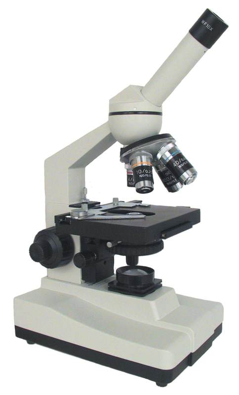
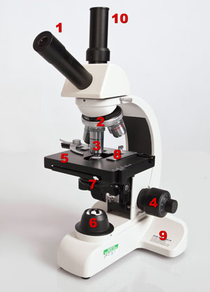
Blank microscope diagram
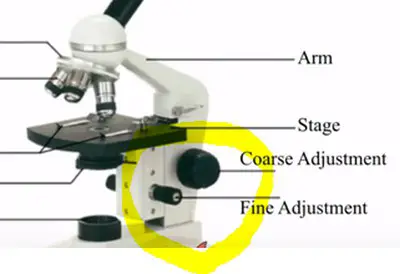
Parts of a Dog - External Body Features of Dogs with Diagram In the dog skeleton labeled diagram, I tried to show you all the bones from the body. This might help you understand the different regions of the body so quickly. I would like to show different external features of a dog again here in a labeled picture. You will find more dog external features labeled diagrams on social media of anatomy learners. Microscope: Definition, Anatomy, Types and Uses - Embibe There microscope anatomy includes three structural parts, i.e. head, base, and arm. Head - This is also known as the body; it carries the optical parts in the upper part of the microscope.. Base - It acts as microscopes support.It also carries microscopic illuminators. Arms - The microscope arm connects the base and the head and the eyepiece tube to the microscope base. Microscope Types (with labeled diagrams) and Functions Has a higher level of magnification - Typically up to 2000x. Is used in hospitals and forensic labs by scientists, biologists and researchers to study micro organisms. Compound microscope labeled diagram. Compound microscope functions: It finds great application in areas of pathology, pedology, forensics etc.
Blank microscope diagram. Skin: Cells, layers and histological features | Kenhub The organ constitutes almost 8-20% of body mass and has a surface area of approximately 1.6 to 1.8 m2, in an adult. It is comprised of three major layers: epidermis, dermis and hypodermis, which contain certain sublayers. Owing to variations in height and weight, the surface area of the skin may vary based on these parameters. 5 Whys Template: Free Download | SafetyCulture Solution: Use a digital inspection app to streamline inventory audits and reporting. Team member 1: Look for digital inspection apps online. Team member 2: Invite process manager to the next meeting. Team member 3: Remind each team member for the next meeting. 5 Whys Template Example: Assigning Actions. Specifications | ECLIPSE Ni-E | Upright Microscopes | Nikon Microscope ... Ni-E Focusing stage type Ni-E Focusing nosepiece type Ni-U; Filter cube turret: 6 filter cubes mountable, High S/N noise terminator mechanism for all turrets Top 14 Best Microscope Slides Reviews (2022) - Screamsorbet LAKWAR. Prime. Slides for microscope with standard dimensions: 1" x 3" (25mm x 75mm) Great set of slides with a wide variety of interesting specimens, for beginner to practice microscopy, to entertain and educate more. 3.
Binocular Microscope Anatomy - Parts and Functions with a Labeled Diagram If you see the light microscope diagram, you will find a different objective lens that attaches to the nose piece. Usually, you will find three to four objective lenses on a light microscope - 4X, 10X, 40X, and 100X. Again, the eyepiece lens has 10X power so you will get a total of 40X, 100X, 400X, and 1000X magnification. The shortest lens ... Compound Microscope - Diagram (Parts labelled), Principle and Uses A compound microscope: Is used to view samples that are not visible to the naked eye. Uses two types of lenses - Objective and ocular lenses. Has a higher level of magnification - Typically up to 2000x. Is used in hospitals and forensic labs by scientists, biologists and researchers to study microorganisms. Microscope Parts, Types & Diagram | What is a Microscope? The essential parts include the head, base, arms, lenses, and lights. In diagrams, one would see the head always located at the top of the microscope while the base is at the bottom. The arms of a ... Compound Microscope Parts Labeled [REJA3G] This is an online quiz called Microscope Parts Diagram This is an online quiz called Microscope Parts Diagram. The main parts of a microscope that are easy to identify include: Head: The upper part of the microscope that houses the optical elements of the unit They explore various websites, label the parts of a microscope on a worksheet, …
14 Best Buy Microscope In 2022: [Latest Updated] - Newportdunesgolf Brand. Emarth. Prime. 【High Magnification】microscope built-in WF10x & WF25x eyepiece and optical lens: 4x, 10x, 40x , rotatable monocular head offers six magnification levels at 40x, 100x, 250x, 400x and 1000x. High Quality Optics can give children improved visual quality and sharp image when in use. 【Easy to Focus & 6 Colorful Filters ... Scanning Electron Microscope (SEM) - Diagram, Working Principle ... Manfred von Ardenne developed the first version of the SEM in 1937. Q5. What is the cost of a scanning electron microscope? The price of a new electron microscope ranges between $80,000 to $10,000,000 and above depending on the customizations, configurations, resolution, components, and brand value. Types of Microscopes: Definition, Working Principle, Diagram ... Checkout JEE MAINS 2022 Question Paper Analysis : Checkout JEE MAINS 2022 Question Paper Analysis : × Download Now PhysicsOpticsTypes Of Microscope Table of ContentsWhat are the Different Types of Microscopes?Simple MicroscopeSimple Microscope DiagramCompound MicroscopeElectron MicroscopeStereo Micr... Barska 50pcs Blank Microscope Slides (AF11636) | Quill.com Order Barska 50pcs Blank Microscope Slides (AF11636) today at Quill.com and get fast shipping. Stack coupons to get free gifts & extra discounts! ... The 50 Blank Microscope Slides w/wooden case is a great start to viewing specimens on a microscope. Each slide has ground edges, is 1mm in thickness, and is 3 inches long and 1 inch wide. 9.25x4 ...
Bright-field microscope (Compound light microscope) - Diagram (Parts ... A few applications of the bright-field microscope include: Used to observe, analyze, and study plant cells. Used to view, magnify, and study about animal cells. Used to clearly study the morphologies of bacterial, and viral organisms. Also used in the study of parasites like paramecium. It finds use in agricultural laboratories to study soil ...
Telomere - Genome.gov Definition. …. A telomere is a region of repetitive DNA sequences at the end of a chromosome. Telomeres protect the ends of chromosomes from becoming frayed or tangled. Each time a cell divides, the telomeres become slightly shorter. Eventually, they become so short that the cell can no longer divide successfully, and the cell dies.
Microscope Types (with labeled diagrams) and Functions Has a higher level of magnification - Typically up to 2000x. Is used in hospitals and forensic labs by scientists, biologists and researchers to study micro organisms. Compound microscope labeled diagram. Compound microscope functions: It finds great application in areas of pathology, pedology, forensics etc.
Microscope: Definition, Anatomy, Types and Uses - Embibe There microscope anatomy includes three structural parts, i.e. head, base, and arm. Head - This is also known as the body; it carries the optical parts in the upper part of the microscope.. Base - It acts as microscopes support.It also carries microscopic illuminators. Arms - The microscope arm connects the base and the head and the eyepiece tube to the microscope base.
Parts of a Dog - External Body Features of Dogs with Diagram In the dog skeleton labeled diagram, I tried to show you all the bones from the body. This might help you understand the different regions of the body so quickly. I would like to show different external features of a dog again here in a labeled picture. You will find more dog external features labeled diagrams on social media of anatomy learners.











Komentar
Posting Komentar