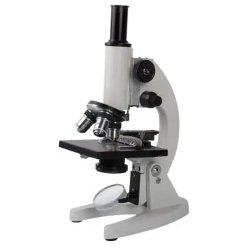
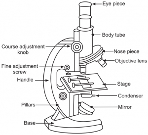
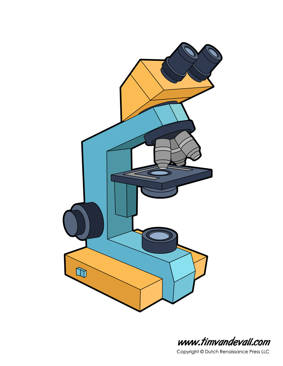
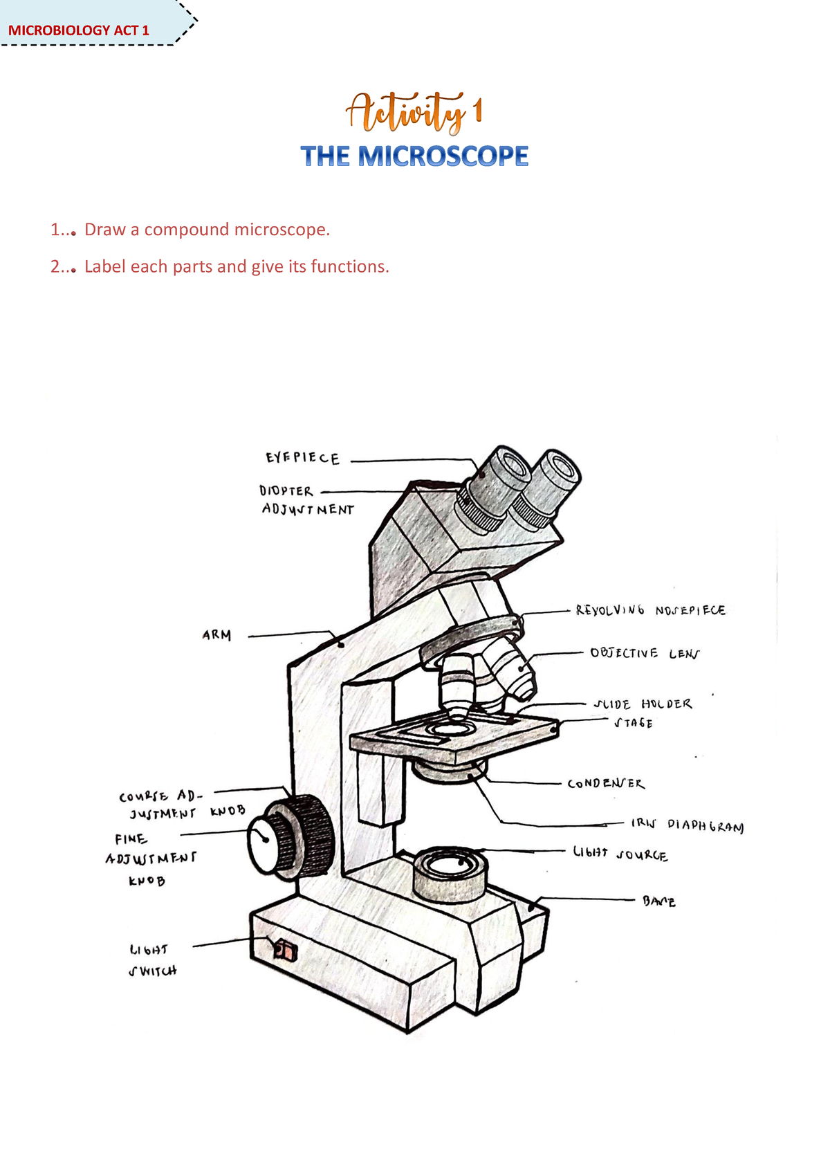
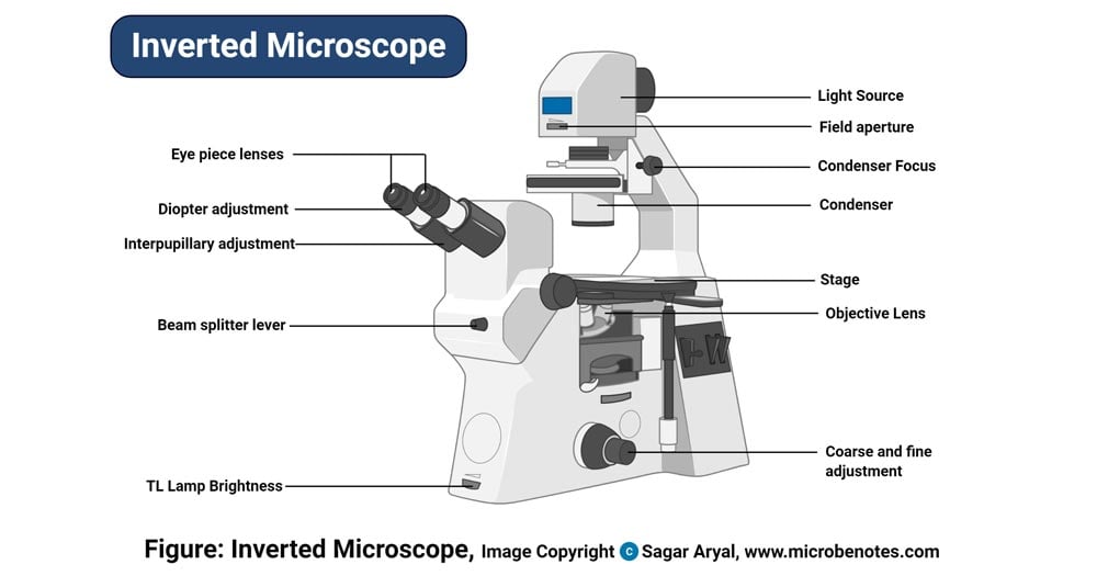
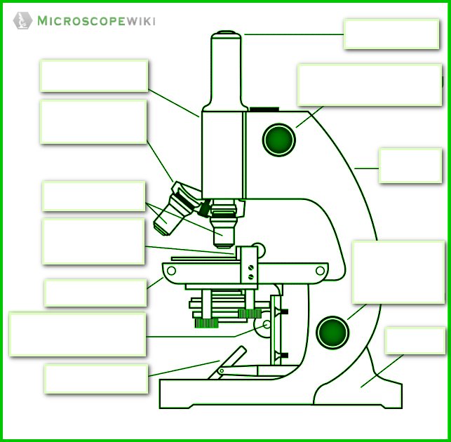
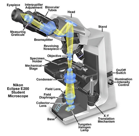
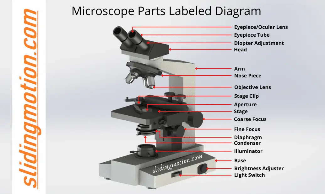
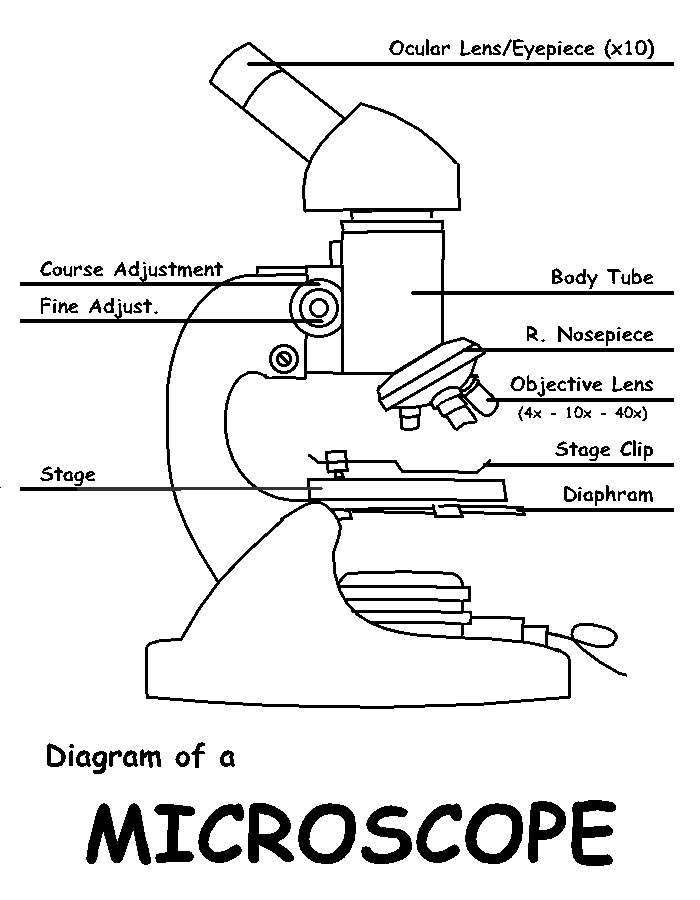
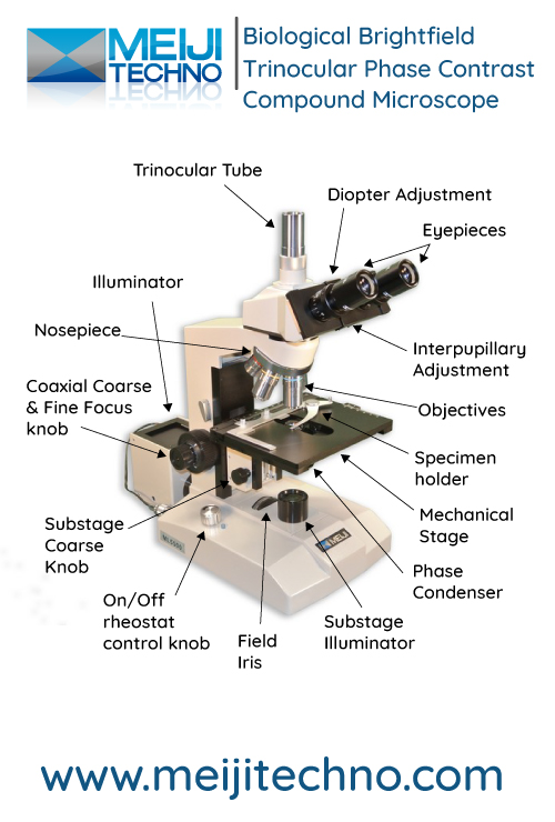
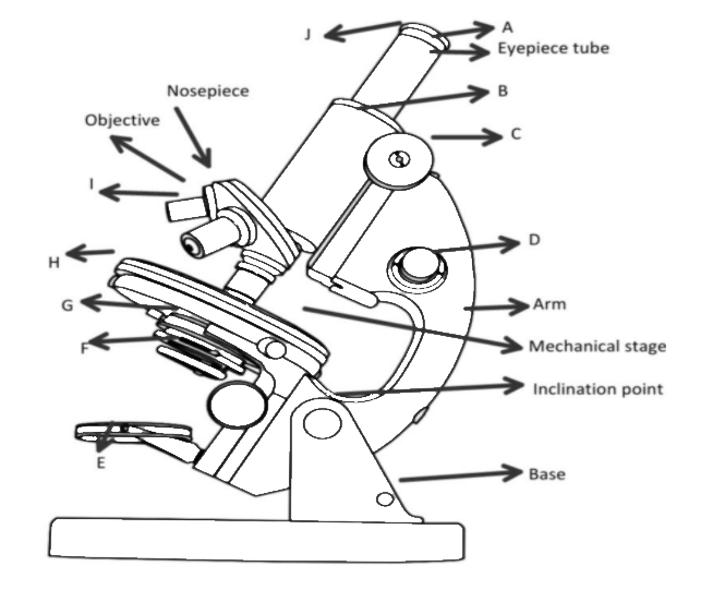
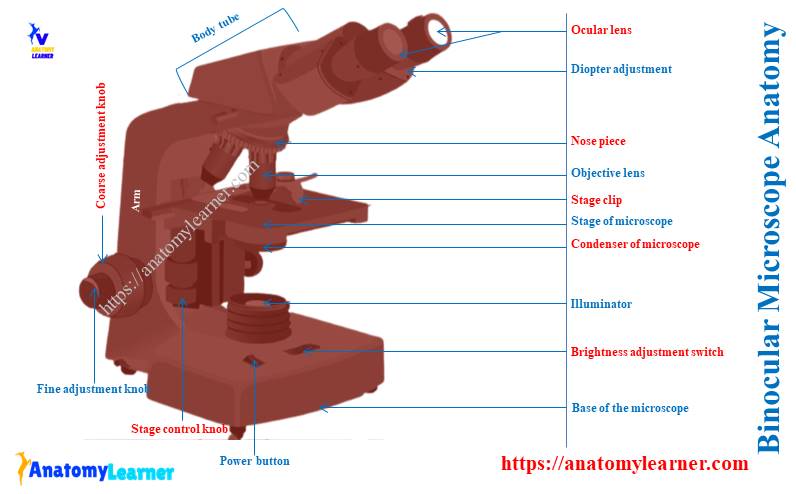
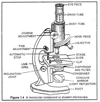
41 labeled diagram of a compound microscope
Compound Microscope Labeled Diagram | Quizlet Compound Microscope Labeled, + −, Flashcards, Learn, Test, Match, Created by, meganplocher734, Terms in this set (14) Eyepiece/Ocular lens, Contains the ocular lens, Body tube, A hollow cylinder that holds the eyepiece. Arm, Part that supports the microscope. Stage, Supports the slide or specimen, Coarse adjustment Knob, Compound microscope- definition, labeled diagram, parts, uses.docx ... View Compound microscope- definition, labeled diagram, parts, uses.docx from BIOLOGY 124 at Harvard University. Compound microscope- definition, labeled diagram, parts, uses October 18, 2018 by Sagar
microbenotes.com › parts-of-a-microscopeParts of a microscope with functions and labeled diagram Apr 19, 2022 · Figure: Diagram of parts of a microscope. There are three structural parts of the microscope i.e. head, base, and arm. Head – This is also known as the body. It carries the optical parts in the upper part of the microscope. Base – It acts as microscopes support. It also carries microscopic illuminators.
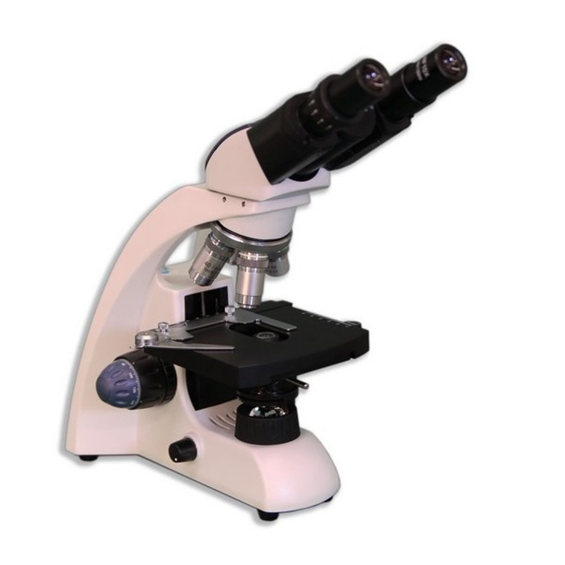
Labeled diagram of a compound microscope
Diagram of a Compound Microscope - Biology Discussion The size of objects viewed under the compound microscope can be accurately determined using a micrometer. The latter consists of two scales, the eyepiece scale, (also called 'graticule' or 'ocular') and the stage micrometer scale. The eyepiece scale is calibrated with the help of stage micrometer and the former is then used for measurements. Compound Microscope Parts - Labeled Diagram and their Functions Labeled diagram of a compound microscope, Major structural parts of a compound microscope, Optical components of a compound microscope, Eyepiece, Eyepiece tube, Objective lenses, Nosepiece, Specimen stage, Coarse and fine focus knobs, Rack stop, Illuminator, Condenser, Abbe condenser, Iris Diaphragm, Condenser Focus Knob, Summary, › ternary-phase-diagramTernary Phase Diagram - an overview | ScienceDirect Topics A point on the diagram represents a composition that is specified in terms of mole fraction or weight fraction. The point, (0.3, 0.4, 0.3) is at the center of the small triangle in the diagram and is located by following the red diagonal 60° line at red 0.3 and the horizontal line at blue 0.4 or any combination of two of the coordinates (A, B, C).
Labeled diagram of a compound microscope. Microscope Types (with labeled diagrams) and Functions A compound microscope: Is used to view samples that are not visible to the naked eye, Uses two types of lenses - Objective and ocular lenses, Has a higher level of magnification - Typically up to 2000x, Is used in hospitals and forensic labs by scientists, biologists and researchers to study micro organisms, Compound microscope labeled diagram, microscopewiki.com › compound-microscopeCompound Microscope – Diagram (Parts labelled), Principle and ... Feb 03, 2022 · See: Labeled Diagram showing differences between compound and simple microscope parts Structural Components. The three structural components include. 1. Head. This is the upper part of the microscope that houses the optical parts. 2. Arm . This part connects the head with the base and provides stability to the microscope. Parts of a Compound Microscope and Their Functions - NotesHippo Compound microscope mechanical parts (Microscope Diagram: 2) include base or foot, pillar, arm, inclination joint, stage, clips, diaphragm, body tube, nose piece, coarse adjustment knob and fine adjustment knob. Base: It's the horseshoe-shaped base structure of microscope. All of the other components of the compound microscope are supported ... Compound Microscope Parts, Functions, and Labeled Diagram Compound Microscope Parts, Functions, and Labeled Diagram, Parts of a Compound Microscope, Each part of the compound microscope serves its own unique function, with each being important to the function of the scope as a whole.
Draw a labelled ray diagram of a compound microscope and ... - Vedantu In a compound microscope the focal length of the objective is 0.5 cm and the focal length of eyepiece is 5 cm. The real image of the object is formed at a distance of 15.5 cm from the objective. If the final image is formed at a distance of 25 cm from the eyepiece, what is the magnifying power of the microscope? A. 180 B. 0.2 C. 20 D. 150 Microscope Parts and Functions Most specimens are mounted on slides, flat rectangles of thin glass. The specimen is placed on the glass and a cover slip is placed over the specimen. This allows the slide to be easily inserted or removed from the microscope. It also allows the specimen to be labeled, transported, and stored without damage. (a) Draw a labelled ray diagram of a compound microscope. (b) Derive an ... (a) Labelled diagram of compound microscope. The objective lens form image A' B' near the first focal point of eyepiece. (b) Angular magnification of objective lens m 0 = linear magnification h'/h. where L is the distance between second focal point of the objective and first focal point of eyepiece.If the final image A'' B'' is formed at the near point. microscopewiki.com › electron-microscopeElectron Microscope Principle, Uses, Types and Images ... Feb 02, 2022 · Ans: A light microscope has a low resolving power (0.25µm to 0.3µm) while the electron microscope has a resolution power about 250 times higher than the light microscope at about 0.001µm. Similarly, a light microscope has a magnification of 500X to 1500x while the electron microscope has a much higher magnification of 100,000X to 300,000X.
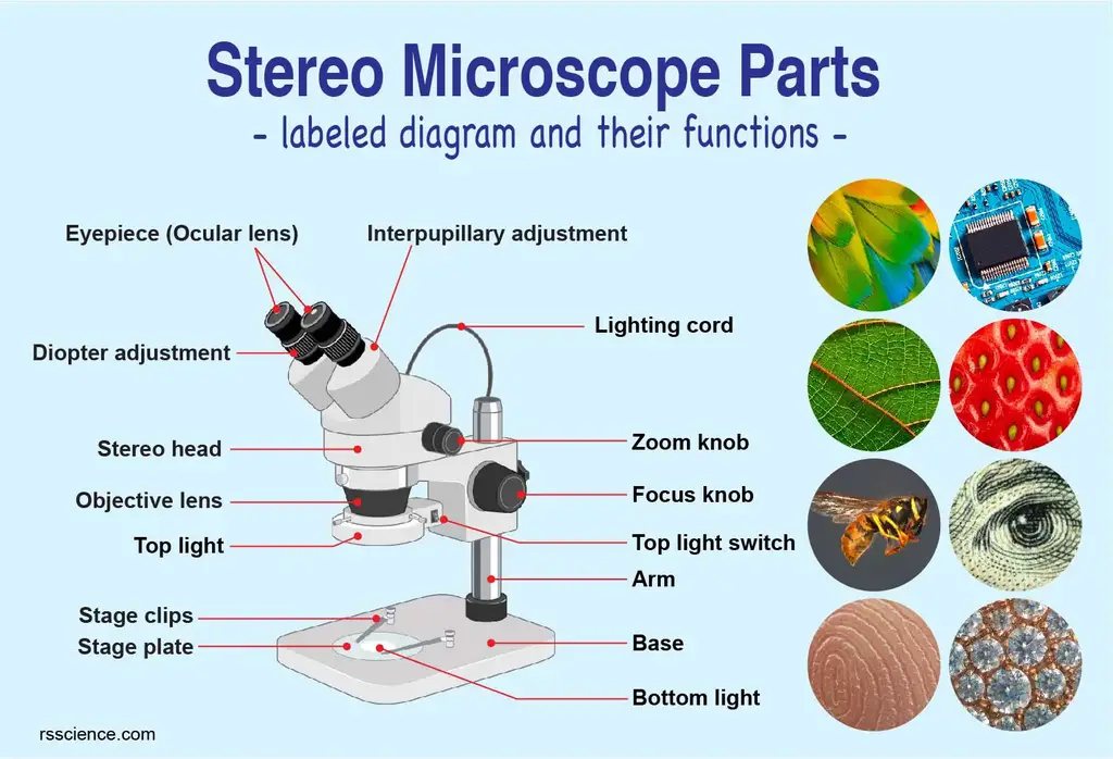
microbenotes.com › compound-microscope-principleCompound Microscope- Definition, Labeled Diagram, Principle ... Apr 03, 2022 · Parts of a Compound Microscope. Eyepiece And Body Tube. The eyepiece is the lens through which the viewer looks to see the specimen. It usually contains a 10X or 15X power lens. The body tube connects the eyepiece to the objective lenses. Objectives and Stage Clips. Objective Lenses are one of the most important parts of a Compound Microscope. Microscope Parts, Function, & Labeled Diagram - slidingmotion Microscope parts labeled diagram gives us all the information about its parts and their position in the microscope. Microscope Parts Labeled Diagram, The principle of the Microscope gives you an exact reason to use it. It works on the 3 principles. Magnification, Resolving Power, Numerical Aperture. Parts of Microscope, Head, Base, Arm, rsscience.com › stereo-microscopeParts of Stereo Microscope (Dissecting microscope) – labeled ... If you would like to learn optical components of a compound microscope, please visit Compound Microscope Parts – Labeled Diagram and their Functions, and this article. How to use a stereo (dissecting) microscope. Follow these steps to put your stereo microscopes in work: 1. Set your microscope on a tabletop or other flat sturdy surface where ... Label a Compound Microscope Diagram | Quizlet Label a Compound Microscope Diagram | Quizlet, Label a Compound Microscope, + −, Flashcards, Learn, Test, Match, Created by, Hesi_Study, Terms in this set (16) Label this, Eyepiece (ocular lens) Label this, Body tube, Label this, Arm, Label this, Mechanical Stage Control Knobs, Label this, Coarse Adjustment Knob, Label this, Fine Adjustment Knob,
A Study of the Microscope and its Functions With a Labeled Diagram ... To better understand the structure and function of a microscope, we need to take a look at the labeled microscope diagrams of the compound and electron microscope. These diagrams clearly explain the functioning of the microscopes along with their respective parts. Man's curiosity has led to great inventions. The microscope is one of them.
Labelled Diagram of Compound Microscope The below mentioned article provides a labelled diagram of compound microscope. Part # 1. The Stand: The stand is made up of a heavy foot which carries a curved inclinable limb or arm bearing the body tube. The foot is generally horse shoe-shaped structure (Fig. 2) which rests on table top or any other surface on which the microscope in kept.
Compound Microscope: Definition, Diagram, Parts, Uses, Working ... - BYJUS The parts of a compound microscope can be classified into two: Non-optical parts, Optical parts, Non-optical parts, Base, The base is also known as the foot which is either U or horseshoe-shaped. It is a metallic structure that supports the entire microscope. Pillar, The connection between the base and the arm are possible through the pillar. Arm,
Microscope, Microscope Parts, Labeled Diagram, and Functions Revolving Nosepiece or Turret: Turret is the part of the microscope that holds two or multiple objective lenses and helps to rotate objective lenses and also helps to easily change power. Objective Lenses: Three are 3 or 4 objective lenses on a microscope. The objective lenses almost always consist of 4x, 10x, 40x and 100x powers. The most common eyepiece lens is 10x and when it coupled with ...
Parts of a Compound Microscope - Labeled (with diagrams) Parts of a Compound Microscope - Labeled (with diagrams) A compound microscope is known as a high-power microscope that enables you to achieve a high level of magnification. Smaller specimens can be thoroughly viewed using a compound microscope. Let us take a look at the different parts of a compound microscope and understand each key component.
microscopeinternational.com › what-is-a-stereoWhat is a Stereo Microscope? - New York Microscope Company May 11, 2018 · A stereo or dissecting microscope is not the same as a compound microscope. Unlike the compound microscope in a stereo microscope, the image is upright and not upside down and backward. A compound microscope yields a single optical path resulting in the same image to both the left and right eye. A stereo microscope provides two optical paths ...
› ternary-phase-diagramTernary Phase Diagram - an overview | ScienceDirect Topics A point on the diagram represents a composition that is specified in terms of mole fraction or weight fraction. The point, (0.3, 0.4, 0.3) is at the center of the small triangle in the diagram and is located by following the red diagonal 60° line at red 0.3 and the horizontal line at blue 0.4 or any combination of two of the coordinates (A, B, C).
Compound Microscope Parts - Labeled Diagram and their Functions Labeled diagram of a compound microscope, Major structural parts of a compound microscope, Optical components of a compound microscope, Eyepiece, Eyepiece tube, Objective lenses, Nosepiece, Specimen stage, Coarse and fine focus knobs, Rack stop, Illuminator, Condenser, Abbe condenser, Iris Diaphragm, Condenser Focus Knob, Summary,
Diagram of a Compound Microscope - Biology Discussion The size of objects viewed under the compound microscope can be accurately determined using a micrometer. The latter consists of two scales, the eyepiece scale, (also called 'graticule' or 'ocular') and the stage micrometer scale. The eyepiece scale is calibrated with the help of stage micrometer and the former is then used for measurements.

















Komentar
Posting Komentar